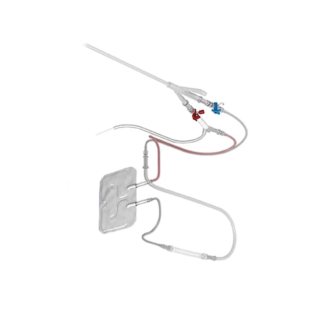
척추 전이 및 기타 골성 병변은 어려운 임상 시나리오를 제시합니다. 통증, 신경학적 손상 및 잠재적인 불안정은 효과적인 치료를 매우 중요하게 만듭니다. 이 골내 고주파 절제(RFA) 시스템은 척추 내 종양 조직의 표적 파괴를 위한 유망, 최소 침습 기술을 제공합니다. 17G 삽입 바늘, 5 Fr 카테터, 다단계 안전 모니터링 기능이 있는 열 절제 프로브를 활용하는 이 시스템은 환자의 안전을 유지하면서 종양 세포를 파괴하도록 제어된 절제를 제공합니다.
주요 기능
<상세정보 id="e-n-accordion-item-2240" 열기>
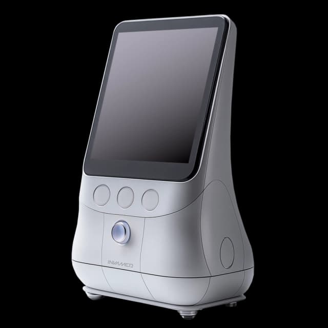
사용자 친화적인 터치스크린 디스플레이
- 직관적인 인터페이스: 작업자는 실시간 피드백을 적극적으로 시각화하는 동시에 절제 매개변수(예: 온도, 전력)를 쉽게 선택하고 미세 조정할 수 있습니다.
- RFID를 통한 프로브 인식: 시스템이 자동으로 카테터 모델을 식별하고 그에 따라 기본 매개변수를 설정하여 간소화 설정을 수행합니다.
실시간 모니터링 및 안전
- 온도 및 전력 출력 추적: 지속적인 분석을 통해 과열 또는 과소 처리를 방지하여 잠재적인 탄화 위험을 제거합니다.
- 경고 시스템: 소리와 빛의 펄스가 높거나 낮은 온도를 신호하고 시술의 대기 작동 시간(최대 250초)을 나타냅니다.
통제된 절제
- 탄화 없음: 40W의 최대 전력으로 정의된 범위(80~110°C)의 온도를 유지하여 과도한 열 확산을 최소화하면서 효과적인 조직 괴사를 가능하게 합니다.
- 다단계 접근 방식: 보다 광범위한 병변의 경우 여러 차례의 순차적 절제를 계획하여 인접 구조와 조직을 보호할 수 있습니다. nerves.
정확한 골내 전달
- 17G 유도관 바늘 + 5Fr 유도관 카테터: 투시경 또는 방사선투시 영상 유도 하에서 척추체에 안정적으로 배치하는 데 도움이 됩니다.
- 열 프로브와 가열 요소(직경 1~2.5mm, 길이 2~4cm): 절제 구역을 병변 크기에 맞게 조정하여 철저한 적용 범위를 보장하고 건강한 뼈 손상을 최소화합니다.
- 직관적인 인터페이스: 작업자는 실시간 피드백을 적극적으로 시각화하는 동시에 절제 매개변수(예: 온도, 전력)를 쉽게 선택하고 미세 조정할 수 있습니다.
- RFID를 통한 프로브 인식: 시스템이 자동으로 카테터 모델을 식별하고 이에 따라 기본 매개변수를 설정하여 간소화 설정을 수행합니다.
- 온도 및 전력 출력 추적: 지속적인 분석을 통해 과열 또는 과소 처리를 방지하여 잠재적인 탄화 위험을 제거합니다.
- 경고 시스템: 소리 및 빛 펄스가 높음 또는 낮음 온도를 신호하고 시술의 대기 작동 시간(최대 25분)을 나타냅니다.0초).
- 탄화 없음: 40W의 최대 전력으로 정의된 범위(80~110°C)의 온도를 유지하여 과도한 열 확산을 최소화하면서 효과적인 조직 괴사를 가능하게 합니다.
- 다단계 접근 방식: 보다 광범위한 병변의 경우 여러 순차적 절제를 계획하여 인접한 구조와 신경을 보호할 수 있습니다.
- 17G 유도 바늘 + 5 Fr 유도 카테터: 투시 또는 방사선 영상 유도 하에서 척추체에 안정적으로 배치할 수 있습니다.
- 열 프로브(가열 기능 포함) 요소(직경 1~2.5mm, 길이 2~4cm): 절제 영역을 병변 크기에 맞게 조정하여 철저한 적용과 건강한 뼈 손상을 최소화합니다.
임상적 장점 및 적용
<상세정보 id="e-n-accordion-item-1440" 열기>
척추 전이 관리
- 통증 완화: 종양 조직의 표적 절제는 통증을 줄이고 손상된 척추뼈의 안정성을 어느 정도 회복할 수 있습니다.
- 최소 침습: 종종 진정제 또는 국소 마취하에 시행되므로 입원 기간이 줄어들고 환자의 이동 속도가 빨라집니다.
합병증 감소 및 보조제 사용
- 과열 위험 감소: 온도 조절을 조절하면 주변 뼈와 척수가 열 손상을 입을 가능성이 줄어듭니다.
- 향상된 시너지 효과: 필요한 경우 절제 후 척추 확대술(예: 시멘트성형술)을 통해 척추를 더욱 안정시킬 수 있습니다.
다양한 이미징 호환성
- 방사선/투시경 유도: 절제 과정에 대한 정확한 탐침 위치 지정과 지속적인 관찰을 보장하여 시술자의 자신감과 환자 안전을 향상시킵니다.
- 통제된 접근 방식: 척추체, 척추경 또는 부속 척추 부위의 전이성 질환에 적합합니다.
절제 후 용이성
- 외부 도입자 없음: 절제 후 단일 17G 액세스 경로를 즉시 철회할 수 있어 절차의 복잡성과 감염 위험을 줄이는 데 도움이 됩니다.
- 즉시 평가: 절차 중 또는 사후 이미징을 통해 절제 영역 범위를 신속하게 확인할 수 있으며 필요한 경우 가능한 2차 절제에 대한 데이터를 제공할 수 있습니다.
- 통증 완화: 종양 조직의 표적 절제를 통해 통증을 줄이고 손상된 척추뼈의 안정성을 어느 정도 회복할 수 있습니다.
- 최소 Inva집중: 진정제 또는 국소 마취 하에 수행되는 경우가 많으며 입원 기간이 줄어들고 환자의 이동 속도가 빨라집니다.
- 과열 위험 감소: 온도 조절을 조절하면 주변 뼈와 척수가 열 손상을 입을 가능성이 줄어듭니다.
- 향상된 시너지 효과: 절제 후, 척추 확대 술(예: 시멘트 성형술)은 필요한 경우 척추를 더욱 안정시킬 수 있습니다.
- 방사선/투시경 유도: 절제 과정에 대한 정확한 프로브 위치 지정과 지속적인 관찰을 보장하여 시술자의 자신감과 환자 안전을 향상시킵니다.
- 통제된 접근 방식: 척추체, 척추경 또는 보조 척추의 전이성 질환에 적합합니다.
- 외부 도입자 없음: 절제 후 단일 17G 액세스 경로를 즉시 철회할 수 있어 절차의 복잡성과 감염 위험을 줄이는 데 도움이 됩니다.
- 즉시 평가: 절차 중 또는 절차 후 영상 촬영을 통해 절제 영역 범위를 신속하게 확인할 수 있으며 필요한 경우 가능한 2차 절제에 대한 데이터를 제공할 수 있습니다.